

What can be found on a pelvic ultrasound?
A pelvic ultrasound is a noninvasive diagnostic test that generates images that are used to evaluate organs and structures in the female pelvis.
The uterus, cervix, vagina, fallopian tubes, and ovaries can all be seen clearly on a pelvic ultrasound.
Ultrasound employs a transducer, which emits ultrasound waves at a frequency that is too high to be heard.
The ultrasound transducer is applied to the skin, and the ultrasound waves travel through the body to the organs and structures.
How does a pelvic ultrasound work?

The ultrasound transducer is applied to the skin, and the ultrasound waves travel through the body to the organs and structures.
The sound waves echo off the organs and return to the transducer.
The reflected waves are processed by the transducer and then converted into an image of the organs or tissues getting analyzed by a computer.
Sound waves travel at various speeds based on the type of tissue encountered, with bone tissue being the fastest and air being the slowest.
The transducer translates the speed at which sound waves are returned to the transducer, as well as the amount of the sound wave returns, as different types of tissue.
Can a pelvic ultrasound detect bowel problems?
Pelvic ultrasound has evolved into a crucial diagnostic tool in the diagnosis of bowel diseases in recent years, owing to technological advances in ultrasonography and increasing physician experience.
When performing a pelvic ultrasound, the sonographer should pay close attention to the bowel to detect GI pelvic diseases such as appendicitis, diverticulitis, colitis, bowel obstruction, mesenteric adenitis, epiploic appendicitis, Crohn’s disease, and even GI malignancy.
What are the limitations of a pelvic scan?
Certain factors or conditions may have an impact on the test results. Among these are, but are not limited to, the following:
- Obesity that is severe
- Barium in the intestines from a recent barium procedure
- Gas in the intestine
- Inadequate bladder filling (with transabdominal ultrasound). For better imaging, a full bladder helps move the uterus up and the bowel away.
How to prepare for a pelvic scan?

Drink at least 700 ml of clear fluid one hour before your appointment. Wait until after the exam to empty your bladder.
Unless the ultrasound is part of another procedure that requires anesthesia, no fasting or sedation is usually required for a pelvic ultrasound.
Your doctor will explain the procedure to you and provide you with the opportunity to ask any questions you may have about it.
Your doctor may request additional preparation based on your medical condition.
Can a pelvic ultrasound detect cancer?
A non-invasive pelvic scan or ultrasound may not be able to detect cancer with efficiency.
However, a transvaginal ultrasound (TVS) test is a scan that uses sound waves to examine the uterus, fallopian tubes, and ovaries by inserting an ultrasound probe into the vagina.
It can aid in the detection of an ovarian mass (tumor), but it cannot determine whether the mass is cancerous or benign.

Pelvic scan for pregnancy
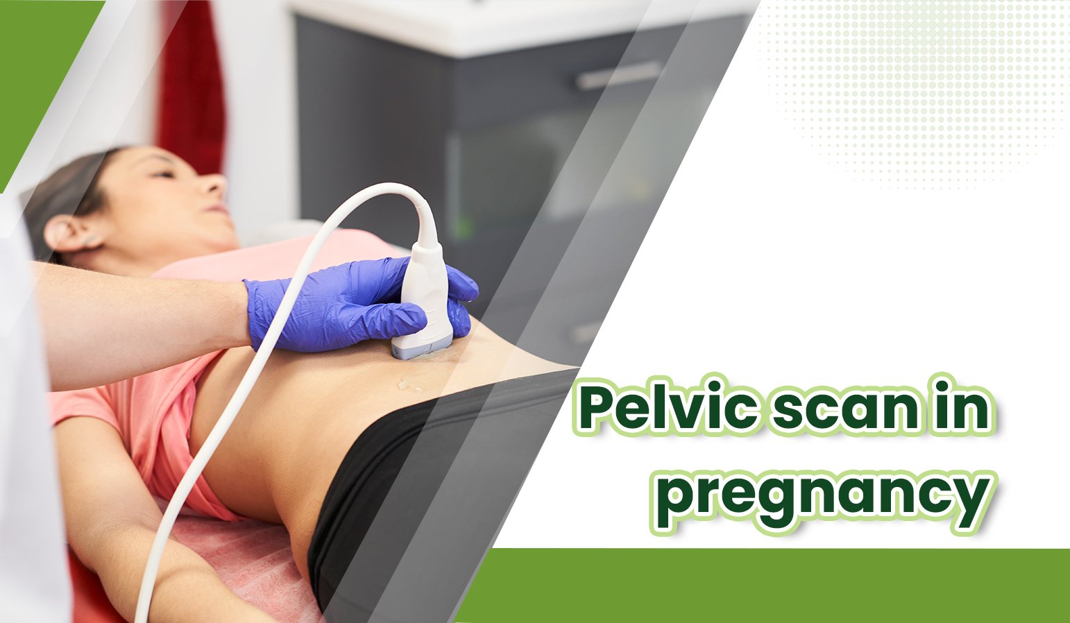
A transabdominal ultrasound is frequently used to track a baby’s development in pregnant women at or before 14 weeks.
The technician will apply a small quantity of warm gel onto one’s stomach and start moving the probe or wand back and forth over one’s stomach for this type of ultrasound.
It will monitor the baby’s growth, including height, arm and leg length, head size, feral heartbeat, placental location and other factors.
It will be used to determine the mother’s pregnancy stage, the baby’s location in the uterus, the number of babies she is carrying, and the volume of amniotic fluid surrounding the baby. It may be used to examine the heart of the baby.
It could be used as a screening test for certain birth defects and congenital malformations, such as Down syndrome, in some instances.
What happens after a pelvic scan?

There is no special care required following a pelvic ultrasound. Unless your doctor advises otherwise, you might very well resume your regular diet and activity.
There have been no substantiated detrimental health impacts on patients or instrument technicians from ultrasound exposure at the levels of intensity used in diagnostic ultrasound.
Depending on your specific situation, your doctor may provide you with added or alternate instructions following the procedure.
