
Growth Scan in Chennai – Third Trimester Pregnancy Ultrasound
Highly accurate fetal growth and well-being assessment performed by fetal medicine specialist Dr. Deepthi Jammi.


Fetal Growth Scan (23–40 Weeks)
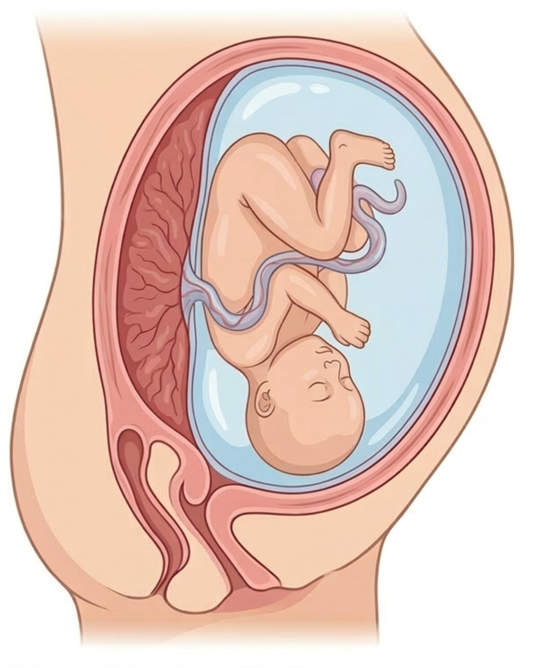
A Fetal Growth Scan, also known as a Growth or Positioning Scan, is a crucial third-trimester ultrasound performed to assess the baby’s growth, development, and overall well-being.
At Jammi Scans, Dr. Deepthi Jammi, a fetal medicine specialist in Chennai, performs all growth scans and provides personal counselling to expectant mothers regarding the scan findings.
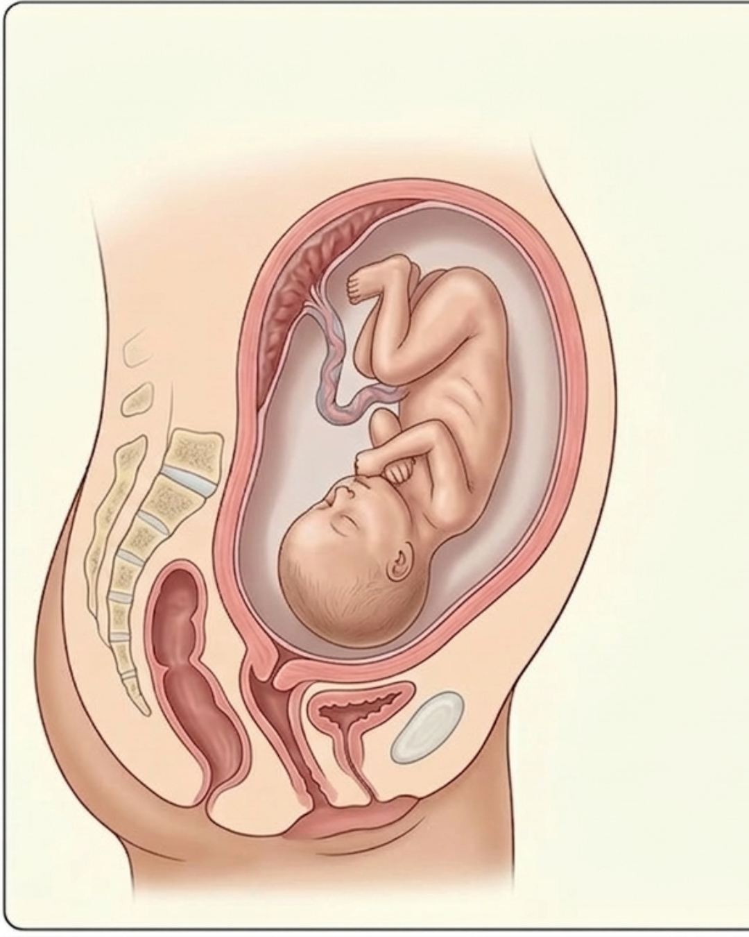
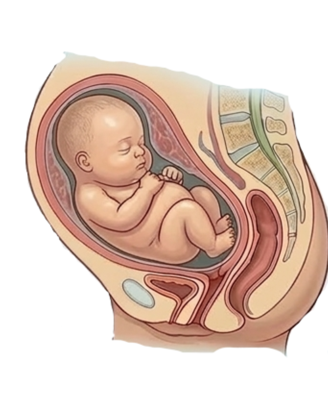
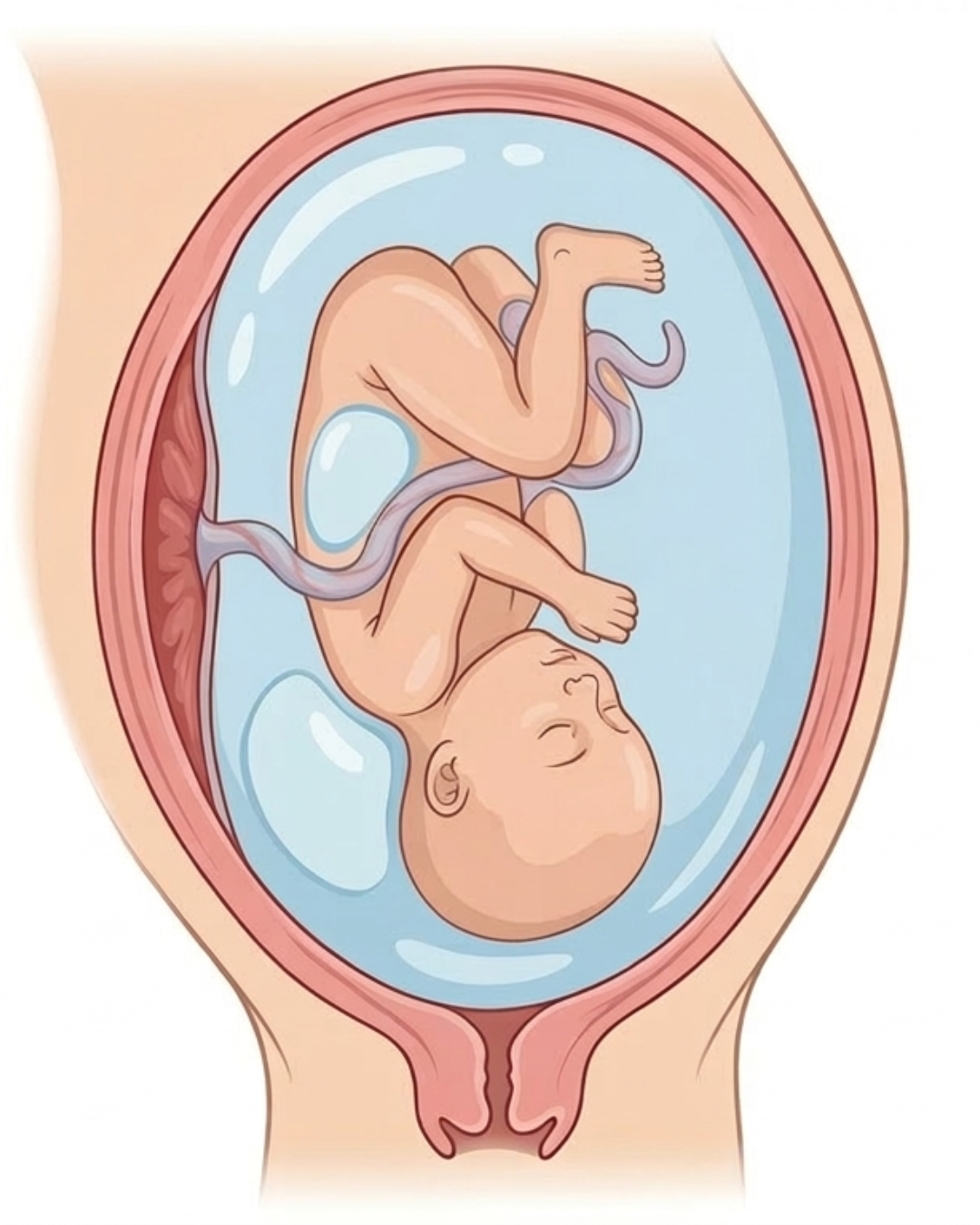
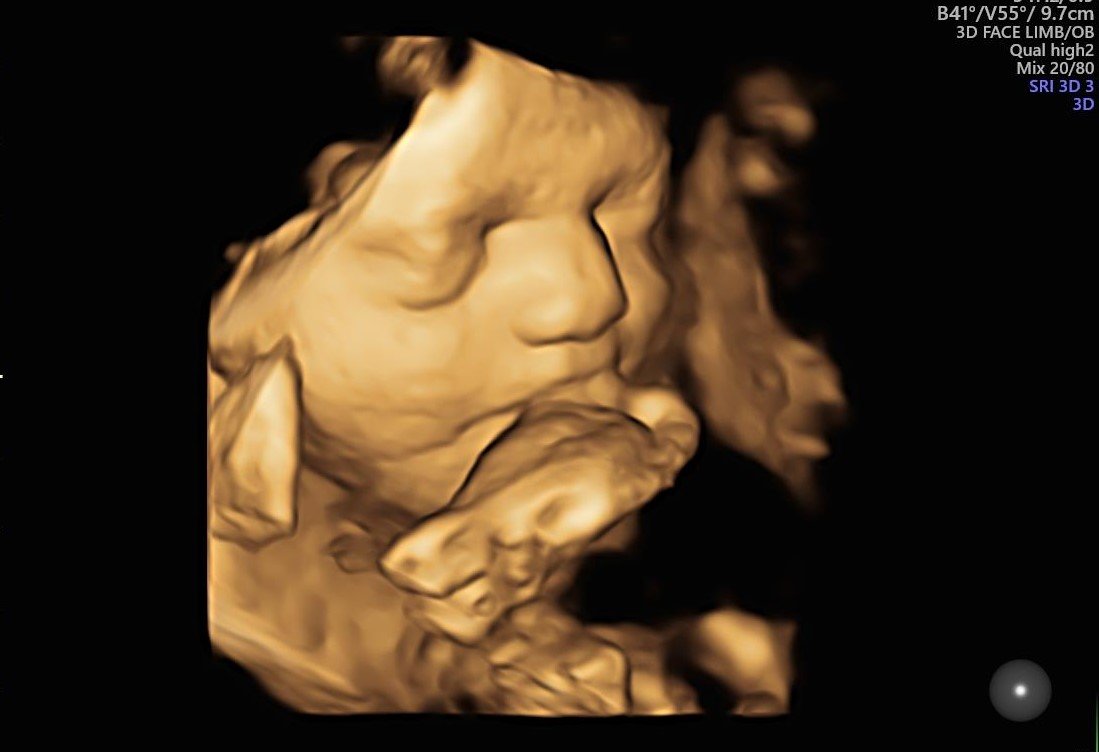 3D image of fetus at 30 Weeks - Jammi Scans
When Should You Do a Growth Scan?
Ideally, a growth scan is performed in the third trimester, between 28 and 40 weeks of pregnancy. In some cases, a growth scan may be repeated if your doctor advises so.
Why Timing Matters
A growth scan is carried out in the third trimester because it is a crucial phase of pregnancy, as the baby grows and develops rapidly during this stage. Growth scans help doctors assess the baby’s growth pattern and plan the birth beforehand.
Accurate Growth Scan in Chennai
A growth scan is carried out to measure the baby’s growth and progress in the later stages of pregnancy. This scan helps assess fetal weight, size, amniotic fluid levels, placental health, and fetal movements to ensure that the baby is developing well for its gestational age.
Growth Scan Report
See what our comprehensive growth scan reports look like. Detailed, accurate, and delivered immediately.
“At Jammi Scans, Dr. Deepthi Jammi provides personal counselling after each scan and guides patients through the next steps.”
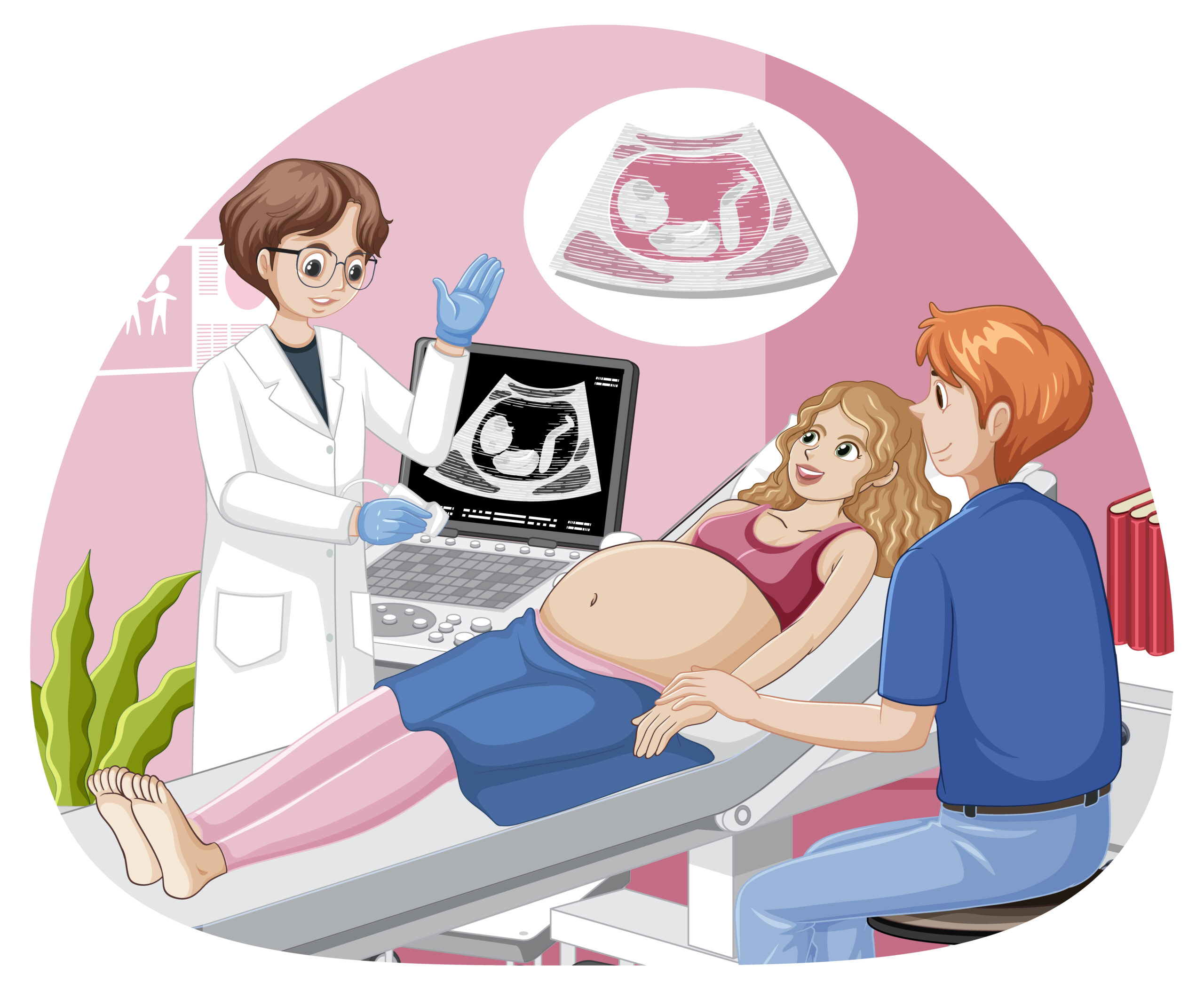
How Is the Growth Scan Performed?
Ideally, a growth scan is done via transabdominal ultrasound.
A thin layer of lubricant gel is applied to the abdomen
The probe (transducer) glides gently over the skin
Real-time images of the fetus are displayed on the monitor
Dr. Deepthi Jammi closely monitors the baby’s biometrics, growth, and development and provides a detailed and accurate report.
Some parents opt for a 3D anomaly scan to see the baby's facial features more clearly.
How to Prepare?

Comfortable Clothing
Wear comfortable clothing.

Full Bladder
Moderately fill your bladder before the scan to improve visibility of the uterus.

No Fasting Required
No special preparation is required before an anomaly scan. You can eat and drink water as usual. There are no dietary restrictions.
What Does a Growth Scan Measure?
The growth scan measures specific fetal parameters, including:
- Biparietal Diameter (BPD) – measures across the head
- Head Circumference (HC) – measures around the head
- Abdominal Circumference (AC) – measures around the abdomen
- Femur Length (FL) – measures the length of the thigh bone
- Estimated Fetal Weight (EFW)
Estimated Fetal Weight (EFW)
Estimated fetal weight (EFW) is the approximate weight of the baby calculated using the biometry measurements.
EFW Percentile Guide
Small for Gestational Age (SGA)
SGA, or Small for Gestational Age, is a term used when the fetus is smaller than other fetuses of the same gestational age.
These babies are smaller than approximately 90% of babies at the exact same stage of pregnancy (falling below the 10th percentile).
SGA can be caused by various factors, including:
- Genetic factors
- Placental issues
- Malnutrition
- Maternal health issues
LGA can be caused by various factors, including:
- Genetics
- Maternal health issues, including gestational diabetes
- The mother being obese or overweight
Large for Gestational Age (LGA)
LGA, or Large for gestational age, is a term used when the fetus is larger than other fetuses of the same gestational age.
These babies are larger than approximately 90% of babies at the exact same stage of pregnancy (falling above the 90th percentile).
Other Observations in a Growth Scan
Apart from measuring the baby’s biometrics, a growth scan also evaluates:
Abnormalities Detected in a Growth Scan
A growth scan is performed in the later stages of pregnancy to monitor the baby’s growth and health. But it also helps rule out certain abnormalities in the fetus including:
-
IUGR (Intrauterine Growth Restriction(1/5))
A condition where the fetus is smaller than average
-
Macrosomia(2/5
A condition where the fetus is larger than average
-
Oligohydramnios(3/5
A condition where the amniotic fluid levels are low
-
Polyhydramnios(4/5
A condition where the amniotic fluid levels are excessive
-
Detection of late-onset anomalies(5/5
Certain abnormalities that develop later in pregnancy, such as skeletal growth abnormalities, fetal growth restriction (FGR), certain heart conditions, and bowel abnormalities
Advanced Growth Scan Options
Comprehensive evaluations combining growth assessment with Doppler or Fetal Echo.
Growth Scan with Doppler
A Growth Scan with Doppler is an advanced third-trimester ultrasound that combines fetal growth assessment with Doppler blood flow studies to evaluate how well blood and oxygen are reaching the baby.
While a standard growth scan measures the baby’s size, weight, and overall development, the Doppler study assesses blood circulation between the mother, placenta, and baby. This helps doctors understand whether the baby is receiving adequate oxygen and nutrients.
When Is it Recommended?
A growth scan with Doppler is commonly advised in cases such as:
- Suspected fetal growth restriction (FGR / IUGR)
- High-risk pregnancies, including hypertension or diabetes
- Reduced fetal movements
- Previous pregnancy complications
- Abnormal growth scan findings
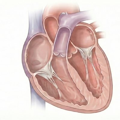
Growth Scan with Echo
While a growth scan is carried out to assess the baby’s growth and development, a fetal echo is carried out to assess the baby’s heart structure and function, helping plan delivery and newborn care if required.
When Is it Recommended?
A growth scan with fetal echocardiography is advised when both the baby’s growth and heart need detailed evaluation, especially in higher-risk pregnancies such as:
- Suspected heart issues
- High-risk maternal conditions such as diabetes, autoimmune disorders, or certain infections
- Growth-related issues in the fetus such as FGR
- Abnormal Doppler findings
- Previous abnormal scan findings, such as increased NT
- IVF or multiple pregnancies
FAQs
Quick answers to your most common questions.
Yes. A growth scan uses ultrasound sound waves and is completely safe for both the mother and the baby.
A growth scan usually takes between 15 and 30 minutes, depending on the baby’s position.
No. A growth scan is a simple, non-invasive ultrasound scan and is not painful.
Not necessarily. The mode of delivery is not dependent solely on the baby’s size. Other factors include the baby’s position, the number of fetuses, and maternal health complications.
While a growth scan and Doppler scan are often carried out together, they are not the same. A growth scan is used to monitor the baby’s growth and development, while a Doppler scan assesses blood flow and oxygen supply to the baby.
At Jammi Scans, growth scan reports are usually provided within 15 minutes.
Yes. At Jammi Scans, attenders are allowed to accompany the mother and share the special moment of seeing the baby.
A growth scan is crucial in analysing the baby’s growth and developmental progress. It allows timely monitoring and intervention if required and helps doctors plan the birth beforehand.
Limitations of a Growth Scan
Estimated fetal weight is not exact
The baby’s weight provided in a growth scan is only an approximation and actual birth weight of the baby may vary.
Image quality may be affected by maternal factors
Factors such as maternal obesity, or reduced amniotic fluid can sometimes limit image clarity and the accuracy of certain measurements.
Labour and delivery events cannot be predicted
A growth scan helps assess the baby’s growth and development, but it cannot predict outcomes such as the exact timing of labour, and mode of delivery.
Important: Despite these limitations, the NT scan remains one of the most valuable tools in prenatal assessment. At Jammi Scans, Dr. Deepthi Jammi uses advanced ultrasound technology and specialised expertise to provide the most accurate evaluation possible.
Why Choose Jammi Scans for Pregnancy Ultrasound?
Specialized care, advanced technology, and compassionate expertise — all focused on your pregnancy journey.

Jammi Scans
At Jammi Scans, all scans are performed by Dr. Deepthi Jammi, an expert Fetal Medicine Specialist, ensuring high standards of accuracy and care for anomaly scans in Chennai.

Reports with in 15 mins
Accurate reports delivered the same day, with clear explanations to help parents understand every detail confidently.

Dr. Deepthi Jammi
Dr. Deepthi Jammi is an FMF-certified fetal medicine specialist (Fetal Medicine Foundation), and she ensures every single FMF protocol is followed to provide accurate results.

Accredited & Certified
Jammi Scans is NABCB-accredited and ISO 9001:2015 certified, meaning we follow the highest standards, undergo regular audits, and ensure that every scan is done precisely.
Pregnancy Ultrasound
Jammi Scans is dedicated exclusively to pregnancy ultrasound and fetal medicine.

Latest Technology
Jammi Scans employs the latest technology ultrasound machines such as the GE Voluson Expert 22 and combines it with Dr. Deepthi Jammi's expertise in fetal medicine to provide comprehensive results.
Pregnancy Ultrasound Scans We Offer
Comprehensive fetal screening at every stage — from confirmation to final growth assessment.
An early pregnancy scan is the first ultrasound scan done to confirm pregnancy. Check the viability of the baby.
- Check the viability of the baby
- Check the baby’s position
- Count the number of babies
- Check the presence of fetal heart rate
- Estimate the baby’s due date
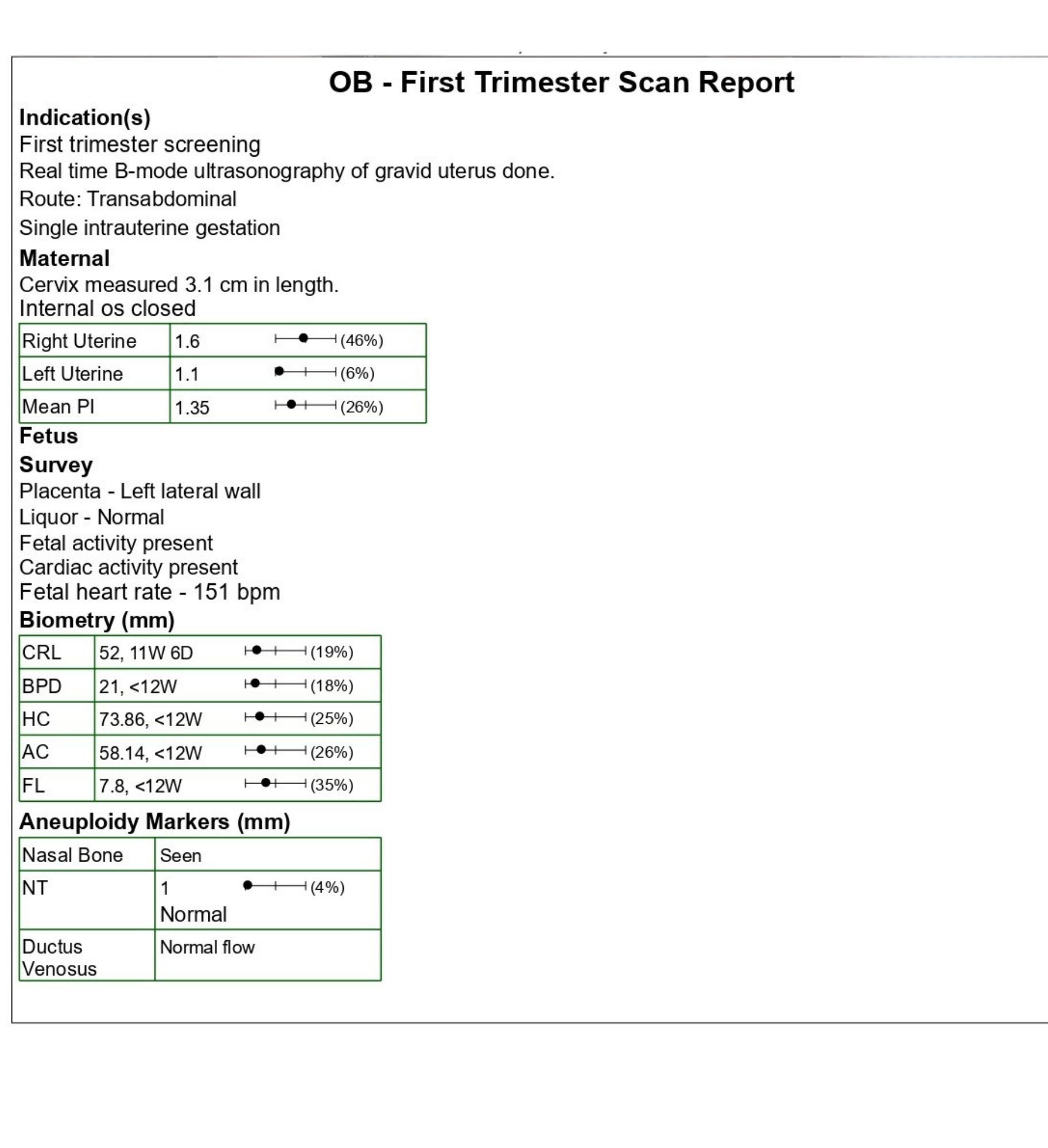
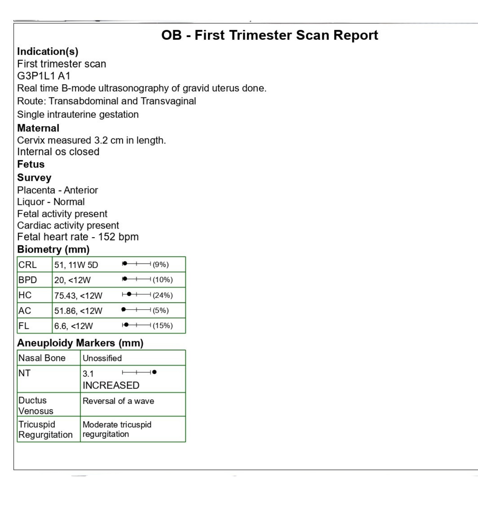
First-trimester screening for chromosomal abnormalities using nuchal translucency (NT) and nasal bone (NB) assessment.
- Down syndrome risk screening
- Fetal nasal bone presence
- Nuchal translucency thickness
- Ductus venosus & tricuspid flow
A detailed anatomy scan is a comprehensive scan used to evaluate fetal development and detect structural anomalies.
- Brain, face, spine & limbs
- Heart (4-chamber view)
- Stomach, kidneys, bladder
- Placenta position & cord
A growth scan is a third-trimester ultrasound that monitors the baby’s growth and development.
- Estimated fetal weight (EFW)
- Amniotic Fluid Index (AFI)
- Fetal position & movements
- Placental maturity
A fetal Doppler scan is used to assess blood flow between the mother, placenta, and baby.
- Umbilical artery PI/RI
- MCA Doppler for anemia risk
- Uterine artery notching
- Combined with Growth Scan
What Our Patients Say
Real reviews from mothers who trusted us with their pregnancy journey.
"I was very nervous about my NT scan, but Dr. Deepthi put me at ease right away. She was very patient and explained everything in a way that I could understand. The scan itself was quick and painless, and the report was very detailed. I would definitely recommend Jammi Scans to anyone."
"I went for my first scan( NT scan) here and the environment is so calm and everything was so organised. They played a video related to the scan and it says why are we doing this scan and what they will check for.Dr.Deepthi mam is very sweet and she will be doing the scan.From my first scan till last scan deepthi mam was there."
"One of the best scan center in Chennai.My wife(gomathi) NT scan done by today. Staffs are very polite and kind.whenever visitors waiting in to main hall is very neat and hygienic. Before scanning proper explanation video's showing and create importance of scan and awareness to each one of you.playing music create to very relaxing mindset.last but not the least finally main core event of scanning by Dr.Deepthi such wonderful person and expert of Handling patient and attender. Very clear explanation to scanning results."
Jammi Scans – Pregnancy Ultrasound Centre, T. Nagar, Chennai
Trusted by 50,000+ mothers for accurate, compassionate, and timely pregnancy scans.
