
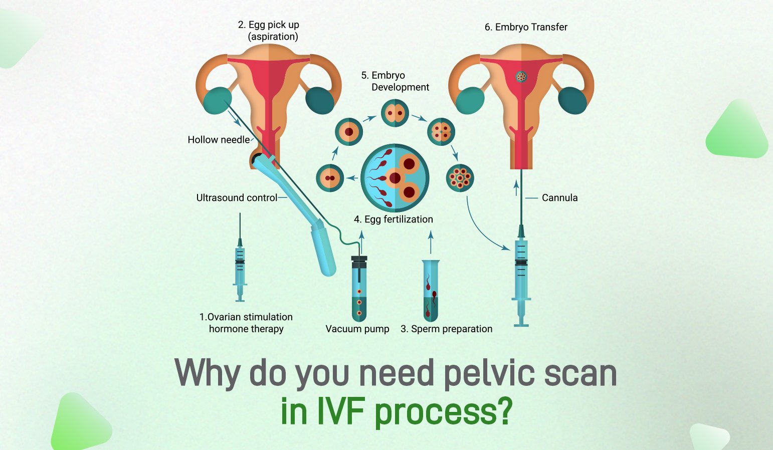
While there are many fertility treatments available that can improve the chance of pregnancy, in vitro fertilisation or IVF, is often considered to be the most versatile and successful. IVF involves retrieving eggs from a woman and then fertilising them with a man’s sperm in a laboratory before transferring the resulting embryos into the woman’s uterus.
Why do you need an ultrasound before IVF?
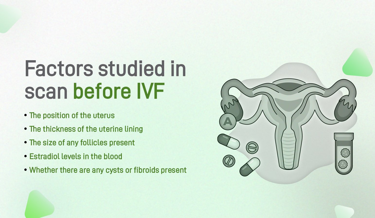
A pelvic scan will be taken at the beginning of IVF treatment to assess the health of the ovaries and check for mature eggs. This examination usually takes place during the woman’s monthly cycle.
Depending on what is found during the pelvic scan, additional treatment may be necessary before IVF can begin. For example, ovarian cysts may require further treatment before proceeding with IVF. However, if everything appears normal, we can proceed with IVF treatment.
What is the scan before IVF?
A baseline ultrasound provides us with important information about the ovaries and pelvic organs, which helps us to determine whether it is the right time to begin ovarian stimulation.
This is the first phase of IVF treatment. A baseline ultrasound may also be performed before transferring frozen embryos.
Can a pelvic scan detect infertility?
In order to determine whether or not a woman is experiencing difficulty in conceiving, it is important to first undergo ultrasound testing. This will verify the presence of the uterus and both ovaries, though it should be noted that ultrasound cannot always reliably detect the fallopian tubes.
In order to properly evaluate this, a hysterosalpingogram may also be performed. This allows for the recording of data such as the size, shape, and position of the uterus, as well as any masses within it (such as fibroids) which can then be mapped.

Additionally, the ovaries can be measured in terms of size and number of follicles present – both of which are important determinants when assessing a woman’s ovarian reserve.
In women, ovarian cysts are commonly found during an infertility ultrasound. Most of the time, these cysts are simply due to the growth of an egg or recent ovulation. However, in some cases, the cyst may be indicative of an abnormality, such as endometriosis.
Additionally, while normal fallopian tubes are not usually seen during an ultrasound, one type of abnormal fallopian tube can be observed. Occasionally, when the distal end of the fallopian tube becomes blocked, it will fill with fluid. This is referred to as hydrosalpinx and can lower the success rate of treatments like IVF.